
Orthognathic surgery
Orthognathic surgery and facial harmonisation: restoring balance and function.
Duration
2 to 5 hours
Anaesthesia
General anaesthesia
Hospital stay
1 to 2 nights
Recovery
Normal activities after 2 to 3 weeks, soft diet for 4 to 6 weeks
A jaw discrepancy is not only about straight teeth: it directly affects chewing, breathing, the health of the temporomandibular joints and the support of the soft tissues of the face.
Misaligned jaws, a receding chin, a protruding jaw, an underbite, a smile showing too much gum or an asymmetric face: when orthodontics alone is not enough, these situations are treated with orthognathic surgery, also called corrective jaw surgery.
Combining advanced maxillofacial surgery with digital technology, orthognathic surgery repositions the jaw bones with millimetre precision to restore a lasting, functional bite and reveal the natural harmony of your profile.
The protocol, step by step.
- Step 1
Orthodontic preparation
The dental arches are aligned for 6 to 18 months before surgery, in close coordination with your orthodontist.
- Step 2
Virtual 3D planning
Merging of the CBCT, the intraoral scans and 3D facial photography, simulation of the bone movements (VSP) and 3D printing of custom surgical splints.
- Step 3
Surgery
Upper and/or lower jaw osteotomies performed from inside the mouth, transfer of the virtual plan to the operating theatre and rigid fixation with plates and screws.
- Step 4
Finishing
Guiding elastics, gentle mouth-opening exercises, post-operative orthodontic adjustments and retention.
More than 25 years’ experience, with patient safety first
Dr Afshin Yousefpour qualified in stomatology in Brussels in 1997, completed full higher surgical training in oral and maxillofacial surgery in the United Kingdom and was appointed Consultant Specialist in 2006. He is a Fellow of the Royal College of Surgeons of Edinburgh (FRCSEd).
In orthognathic surgery, his aim is to restore facial function and harmony while keeping risks and recovery to a minimum. To do so, he relies on virtual 3D planning and, where indicated, piezosurgery (ultrasound), which cuts bone selectively while protecting nerves and soft tissues.
Teamwork with your orthodontist
Orthognathic surgery is a treatment carried out by two specialists. The orthodontist prepares the dental arches before surgery, the surgeon repositions the jaws, then the orthodontist completes the finishing. Dr Yousefpour works closely with referring orthodontists at every stage: initial assessment, timing of surgery, planning and post-operative follow-up.
Virtual 3D planning: digital precision
Data fusion: the cone beam CT scan (CBCT, for bone and nerve anatomy), the intraoral scans (precise bite) and 3D facial photography (to anticipate how the soft tissues and lips will respond).
Virtual surgical planning (VSP): 3D simulation of the bone movements, optimisation of the upper airway and custom 3D-printed surgical splints, to transfer the virtual plan to the operating theatre with full accuracy.
Dr Yousefpour’s particular expertise: complete command of the digital workflow and of advanced 3D simulation software, aligning the bite, skeletal symmetry and soft-tissue dynamics to the millimetre.
Le Fort I osteotomy of the upper jaw
A Le Fort I osteotomy frees the upper jaw so that it can be repositioned in all three planes: forward, upward, downward or to correct an asymmetry.
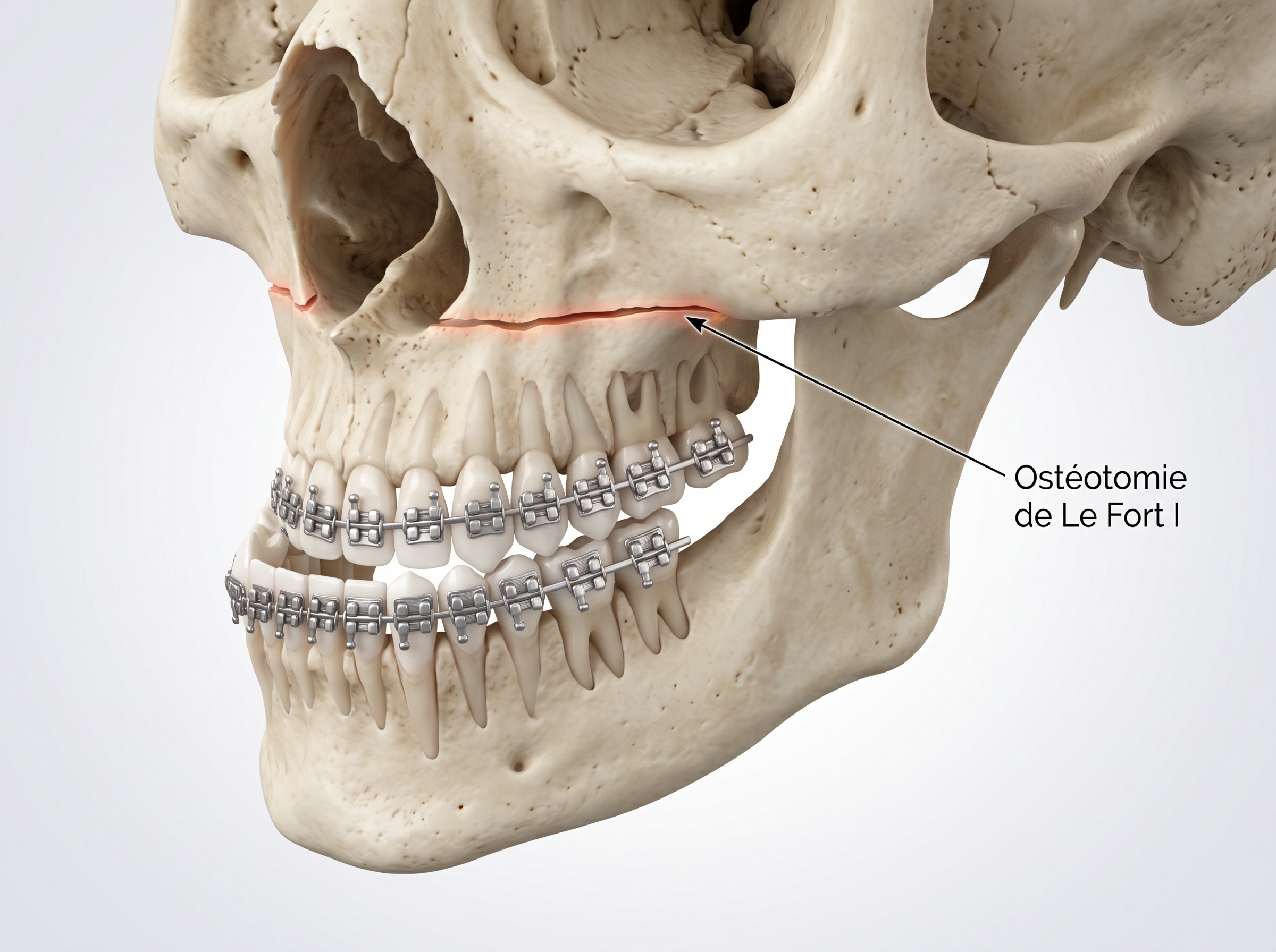
Indications: anterior open bite (front teeth that do not meet), vertical maxillary excess (gummy smile), a retruded upper jaw (flat, sunken profile) or facial asymmetry.
Particular expertise: piezoelectric (ultrasonic) osteotomy for very fine bone cuts that spare the sinus lining and blood vessels, combined with 3D guidance for precise vertical positioning of the midface without affecting the appearance of the nose.
Recovery: marked swelling of the upper jaw and lips for 7 to 10 days, nasal congestion common in the first week, soft or blended diet for 4 to 6 weeks, normal activities after 2 to 3 weeks.
Possible complications: nosebleed, temporary numbness of the upper lip and sides of the nose, post-operative sinusitis, delayed bone healing.
Lower jaw osteotomy (bilateral sagittal split osteotomy)
This technique moves the lower jaw forward, backward or into alignment through a sagittal split of the vertical branches of the jaw, without affecting the dental arch itself.
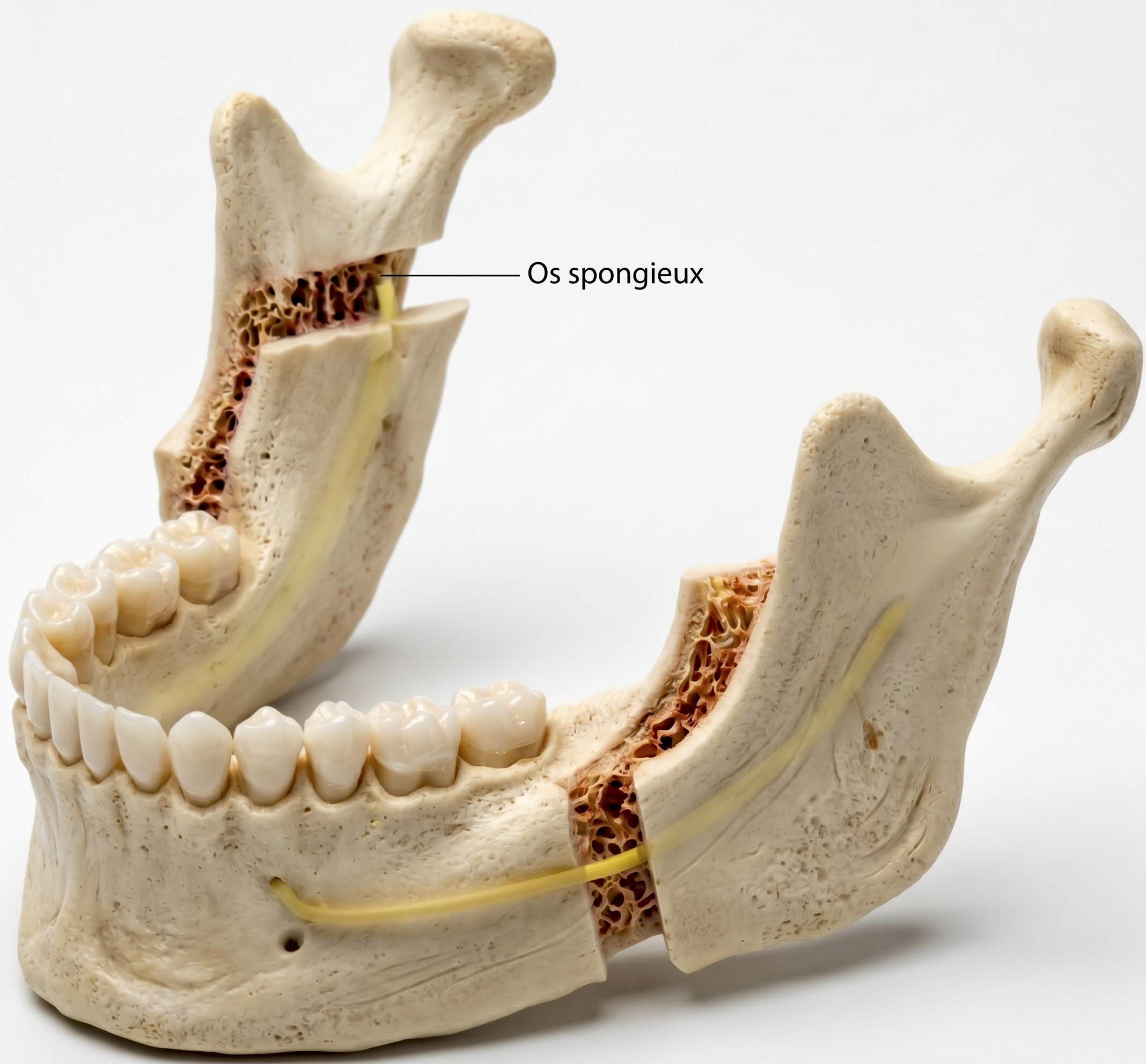
Indications: retrognathia (lower jaw set back, receding chin, often associated with sleep apnoea), prognathism (protruding lower jaw, underbite) and laterognathia (jaw shifted to one side, deviated chin).
Particular expertise: careful 3D mapping of the inferior alveolar nerve on the CBCT and piezoelectric (ultrasonic) osteotomy, a latest-generation technique that cuts bone selectively to minimise the risk of nerve injury and altered sensation, while allowing the jaw joints to settle naturally.
Recovery: swelling of the cheeks and lower face, sometimes bruising on the neck, temporarily limited mouth opening that improves with gentle exercises, light guiding elastics without wiring the jaws shut, liquid then soft diet for 6 weeks.
Possible complications: reduced or altered sensation from the inferior alveolar nerve (temporary numbness of the lower lip and chin, usually recovering over several months; partial lasting numbness is possible but uncommon), delayed bone healing, temporary jaw joint discomfort.
Transverse expansion and palatal distraction (DOME / SARPE)
In adults, the bony sutures of the palate are closed. Surgically assisted maxillary expansion widens the palate in a stable way by combining a minimally invasive osteotomy with a mechanical palatal expander.
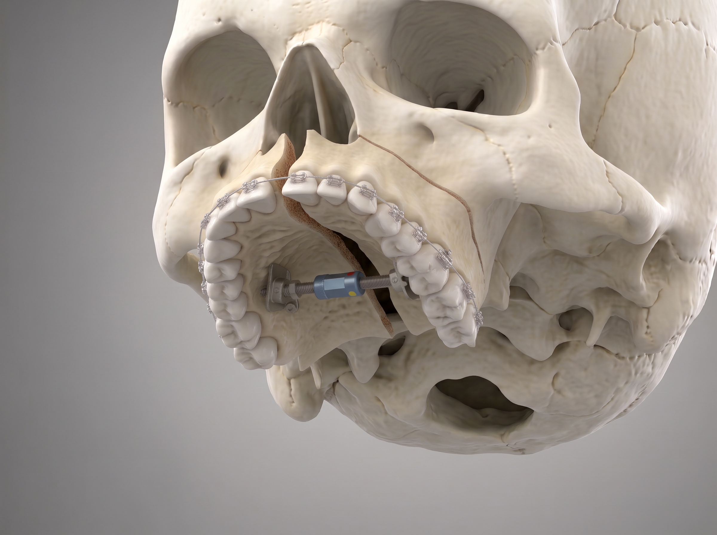
Indications: narrow palate (unilateral or bilateral crossbite), severe crowding, increased nasal resistance and obstructive sleep apnoea.
Particular expertise: targeted, minimally invasive corticotomies and release of the pterygomaxillary junction under direct vision, for parallel, symmetrical expansion without tipping the teeth outwards and an immediate improvement in nasal breathing.
Recovery: moderate discomfort in the palate and a feeling of tension in the nose when the expander is activated each day for 10 to 15 days, a temporary gap between the central incisors that the orthodontist then closes, soft diet during the activation phase.
Possible complications: asymmetric tooth movement, localised gum damage, discomfort from the appliance, partial relapse of the expansion.
Miniscrew-assisted maxillary expansion (MARPE)
MARPE (Miniscrew-Assisted Rapid Palatal Expansion) uses an expander anchored directly in the palatal bone with temporary miniscrews, so that the bone itself is widened while the gums and tooth roots are protected.
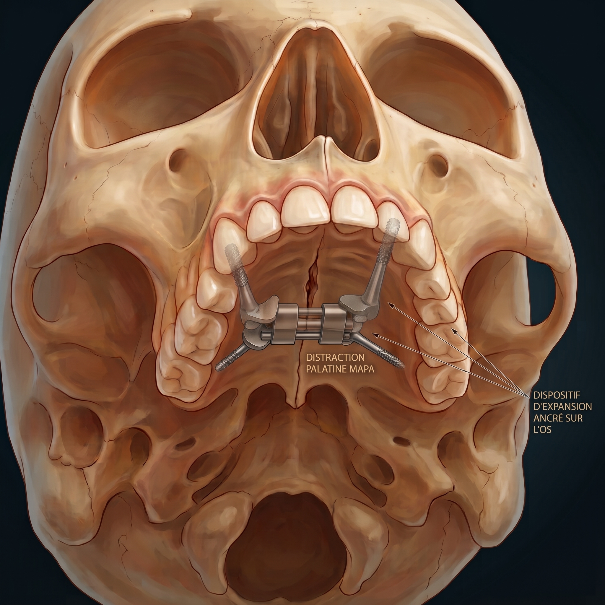
Indications: transverse maxillary deficiency in adolescents at the end of growth or in young adults, as a minimally invasive alternative or complement.
Particular expertise: personalised digital 3D planning of miniscrew placement, based on bone thickness measured on the CBCT, for maximum stability of the appliance and full transmission of the expansion forces to the maxillary skeleton.
Recovery: straightforward, mild pain well controlled by ordinary painkillers, a stretching feeling at the root of the nose, no time off needed.
Possible complications: loosening of, or inflammation around, a miniscrew; in older adults, failure of the midline suture to open, requiring additional surgical assistance.
Functional and aesthetic genioplasty
A genioplasty is an osteotomy of the chin bone that moves the chin in any direction: forward, backward, down, up or towards the midline.
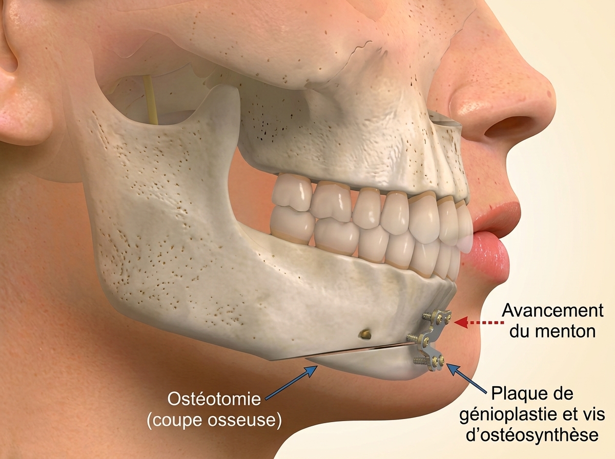
Indications: receding or weak chin, prominent chin, chin asymmetry or lip incompetence (difficulty closing the lips at rest without straining the chin muscles).
Particular expertise: combined analysis of the bony profile and soft tissues using high-fidelity 3D morphing, with custom rigid fixation, to redefine the jawline, better support the muscles under the chin and allow the lips to close naturally, without strain.
Recovery: moderate swelling of the lower face and a pressure dressing for a few days, temporary numbness of the lower lip and chin for a few weeks, return to work after 7 to 10 days.
Possible complications: lasting reduced sensation from the mental nerve, residual asymmetry, bone resorption along the cut lines.
Informed consent: a shared medical decision
Orthognathic surgery is part of a combined medical and surgical treatment in which the maxillofacial surgeon and your orthodontist work closely together.
Informed consent is a fundamental step in your care. Before any operation, a dedicated consultation explains transparently:
The expected functional, breathing and aesthetic benefits.
The stages of digital 3D planning and exactly how the operation will proceed, with the virtual simulations.
The recovery, pain relief and dietary advice.
The surgical risks specific to your anatomy.
Time to reflect, guaranteed
You receive a detailed, personalised consent document, giving you all the time you need to reflect before starting your treatment with confidence.
Frequently asked questions
When is orthognathic surgery indicated?
When a jaw discrepancy cannot be corrected by orthodontics alone: a lower jaw set too far back or too far forward, an open bite, a gummy smile, facial asymmetry, a narrow palate or sleep apnoea related to the position of the jaws.
My jaws are misaligned: can orthodontics alone be enough?
When only the teeth are out of position, orthodontic treatment may be enough. When the jaw bones themselves are misaligned (a receding jaw, a protruding jaw, a jaw shifted to one side), orthodontics can only compensate by tilting the teeth: orthognathic surgery then repositions the jaws, together with your orthodontist. An assessment at the practice (CBCT, intraoral scan and 3D photography) settles the question.
How is a receding chin corrected?
It depends on how the teeth fit together. If the bite is correct, a genioplasty (moving the chin bone) is enough to reshape the profile. If the receding chin comes with a lower jaw set back and upper teeth too far forward (class II), a mandibular advancement corrects the profile, the bite and often night-time breathing at the same time.
My lower jaw sticks out: what can be done?
A protruding lower jaw with the lower teeth in front of the upper teeth (underbite, class III or prognathism) is corrected by orthognathic surgery: moving the lower jaw back, the upper jaw forward or, most often, both (bimaxillary surgery). 3D planning determines which combination best balances the profile and the bite.
Do I need braces before the operation?
Yes, in most cases. Six to eighteen months of orthodontic preparation align the dental arches before surgery, in close coordination between your orthodontist and Dr Yousefpour, followed by orthodontic finishing after the operation.
How is the operation planned?
The CBCT scan, the intraoral scan and 3D facial photography are merged to simulate the bone movements. Custom 3D-printed surgical splints then reproduce this plan precisely in the operating theatre.
How is the risk to the nerve reduced?
The course of the inferior alveolar nerve is mapped in 3D on the CBCT, and piezoelectric (ultrasonic) osteotomy cuts bone selectively to reduce the risk of injury. Temporary numbness of the lip remains possible; it usually recovers within a few months.
How long are the hospital stay and recovery?
The operation takes 2 to 5 hours under general anaesthesia, with 1 to 2 nights in hospital. Normal activities resume after 2 to 3 weeks, with a soft diet for 4 to 6 weeks.
Is orthognathic surgery reimbursed?
As a functional procedure, orthognathic surgery qualifies for reimbursement by the Belgian health insurance funds (mutualités). The costs are given to you in writing before any decision.
Before and after photos: orthognathic surgery
Two examples of orthognathic surgery performed by Dr Yousefpour. The first patient had bimaxillary surgery: a Le Fort I osteotomy to advance the upper jaw, combined with a Dal Pont sagittal split osteotomy to set the lower jaw back, correcting her class III malocclusion. The second patient had a Dal Pont sagittal split osteotomy to advance the lower jaw, correcting a class II malocclusion with a retruded mandible. Every result is specific to the patient and their anatomy and is not a guarantee.
Two patients shown. Tap or click a photo to enlarge it.